All testing is done in our Accredited onsite facility:
Our Nuclear, Echo, and Vascular labs are all ACCREDITED
• Cardiovascular Consultations
• Physicals
• Nuclear Myocardial Perfusion Stress Test ( Exercise)
• 24 Hour Holter Monitoring
• Stress Test ( Pharmacologic)
• Exercise Treadmill Stress Testing
• Echocardiogram
• Stress Echo
• Remote Ambulatory Blood Pressure Monitoring
• Pacemaker/Defibrillator Monitoring
• Coumadin Therapy Monitoring
• Vascular Testing Carotid Duplex
• Renal artery Duplex
• Abdomional aortic duplex
• Lower Extremity venous duplex
• Pulse Volume Recording (PVR)
ILR: Implantable Loop Recorder (or insertable cardiac monitor).


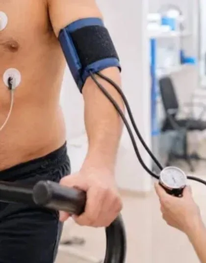
Nuclear and Exercise Stress Testing
A nuclear stress test (myocardial perfusion imaging) evaluates blood flow to the heart muscle at rest and during stress. It uses a small amount of radioactive tracer injected into a vein and a special camera to detect how well blood reaches different areas of the heart.
Why it’s ordered:
To diagnose coronary artery disease (blockages in the heart’s arteries).
To assess the severity and location of ischemia (areas with poor blood flow).
To guide treatment decisions: medical therapy, angiography, or revascularization (stent or bypass).
To evaluate symptoms like chest pain, shortness of breath, or abnormal ECG findings.
To estimate prognosis and risk of heart attack or cardiac events.
Common types:
Exercise stress test (treadmill)
Stress Echocardiography with exercise
Nuclear (radionuclide) stress test with exercise
Pharmacologic (chemical) stress testing without exercise. Used when a patient cannot exercise adequately. Drugs that increase heart rate or dilate coronary vessels are given, always combined with imaging.
Bottom line:
Nuclear stress testing is a well-established, reliable method to evaluate blood flow to the heart under stress and at rest. It helps diagnose coronary artery disease, estimate risk, and guide treatment decisions. Discuss the expected benefits, alternatives, and any concerns with your cardiology team to choose the right test for your situation.
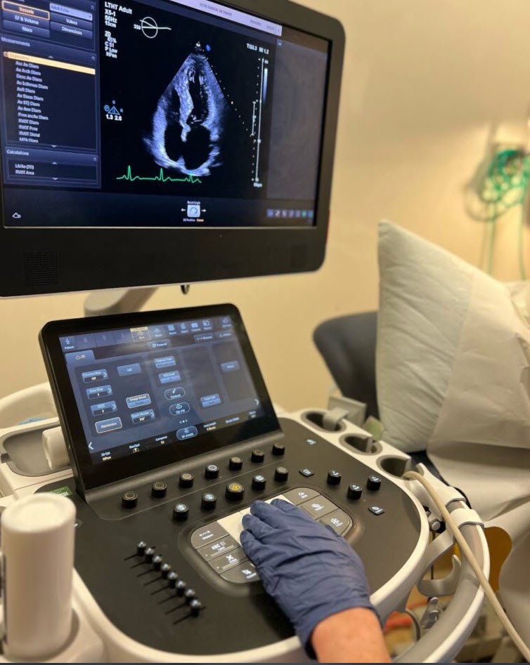
Echocardiogram and Vascular Testing
An echocardiogram (echo) is a noninvasive ultrasound test that uses sound waves to create moving images of your heart. It shows heart structure (chambers, valves, walls) and heart function (pumping strength, blood flow patterns).
Why it’s ordered:
Evaluate symptoms: shortness of breath, chest pain, palpitations, syncope.
Diagnose and monitor: heart failure, valve disease (stenosis, regurgitation), congenital heart defects, pericardial disease, cardiomyopathies.
Assess function: ejection fraction, wall motion abnormalities after a heart attack.
Guide procedures: valve repair/replacement planning, device placement, intraoperative monitoring.
Common types:
Transthoracic echocardiogram (TTE): Standard, performed with a transducer on the chest wall. Quick and painless.
Stress echocardiogram: Echo images are taken before and after exercise (or pharmacologic stress) to detect ischemia or exercise-induced valve problems.
Doppler echocardiography: Measures blood flow direction and velocity to assess valve function and pressure gradients.
Bottom line:
Echocardiography is a first-line, versatile, safe imaging tool for evaluating heart structure and function. Most echoes are simple and painless; some (TEE, stress echo) require special preparation and monitoring. Results guide diagnosis and treatment decisions for a wide range of cardiac conditions.

Cardiology consultations and expert heart care
Common reasons you need a cardiology consultation
Chest pain or pressure: Any new, unexplained, or concerning chest discomfort should be assessed to rule out heart attack, unstable angina, or other cardiac causes.
Shortness of breath: Breathlessness with exertion, at rest, or with fluid buildup can signal heart failure, valvular disease, or ischemia.
Palpitations or irregular heartbeat: Fast, slow, skipped, or irregular beats may indicate arrhythmias that need diagnosis and treatment.
High blood pressure that’s hard to control: Resistant or severe hypertension sometimes has cardiac complications or secondary causes that require specialist input.
Heart murmurs: New or changing murmurs can reflect valvular disease that needs evaluation by a cardiologist.
History of heart attack, stent, bypass, or heart failure: Ongoing specialist care improves outcomes, medication management, and surveillance.
Fainting or near-fainting (syncope): This can be caused by dangerous arrhythmias or structural heart disease.
Pre-operative clearance for major surgery: Patients with cardiac risk factors often need cardiology assessment before elective operations.
Family history of inherited cardiac conditions: Genetic or familial heart disease (e.g., hypertrophic cardiomyopathy, sudden cardiac death) may warrant specialist screening.
Bottom line:
If you have concerning symptoms, abnormal heart tests, a significant cardiac history, or risk factors that aren’t responding to initial treatment, see a cardiologist — sooner rather than later. Your heart will thank you (probably with more regular beats and fewer dramatic plot twists).